Home
/ Surface Anatomy Of Ribs, Pin on anatomy - All valves are a bit lower when the patient is standing.
Surface Anatomy Of Ribs, Pin on anatomy - All valves are a bit lower when the patient is standing.
Surface Anatomy Of Ribs, Pin on anatomy - All valves are a bit lower when the patient is standing.. Ribs the ribs partially enclose and protect the chest cavity, where many vital organs (including the heart and the lungs) are located. Surface markings of the abdomen bony landmarks.—above, the chief bony markings are the xiphoid process, the lower six costal cartilages, and the anterior ends of the lower six ribs. The rib cage surrounds the lungs and the heart, serving as an important means of bony protection for these vital organs. It is located at the level of intervertebral disc between the t4 and t5 vertebrae. Retrospective assessment of hrct examinations of 250 patients was performed.
Retrospective assessment of hrct examinations of 250 patients was performed. An articular capsule surrounds the head of each rib and is further. The uppermost visible digitation of serratus anterior indicates the sixth rib. The ribs are curved, flat bones which form the majority of the thoracic cage. Each rib consists of a head, neck, and a shaft.
The Bones of the Thorax - the rib cage | Anatomy bones ... from i.pinimg.com However, only seven have a direct articulation with the sternum. The first seven sets of ribs, known as true ribs also known as vertebrosternal ribs, are directly articulate with the vertebral column posteriorly and terminate anteriorly as costal cartilage. Contributing to their role in protecting the internal thoracic organs. Size of body l3 c2 apex of co. The lower border of the pectoralis major at its attachment corresponds to the fifth rib; The line diverges somewhat as it descends, and lateral to it is a broad convex surface caused by the projection of the ribs beyond their angles. The upper edge is round and the lower sharp. The lung is located deep to the area going from axilla to the level of the 7th or 8th rib.
All valves are a bit lower when the patient is standing.
There are twelve (12) pairs of ribs and all articulate posteriorly with the thoracic vertebrae. Free for commercial use high quality. It is located at the level of intervertebral disc between the t4 and t5 vertebrae. An articular capsule surrounds the head of each rib and is further. Free for commercial use no attribution required high quality images. The first seven sets of ribs, known as true ribs also known as vertebrosternal ribs, are directly articulate with the vertebral column posteriorly and terminate anteriorly as costal cartilage. Count the ribs and intercostal spaces. The typical rib consists of a head neck and body. Surface projections of the major organs of the trunk, using the vertebral column and rib cage as main reference points of surface anatomy. They are extremely light, but highly resilient; All valves are a bit lower when the patient is standing. The ribs are a set of 12 pairs bones which form the protective 'cage' of the thorax. With the upper ribs, closer to the nodule (and in the case of lower ribs, a little further from the nodule) they are curved and have a rough surface that connects them with muscles, angulus costae.
Because rib 1 articulates with first thoracic vertebra only, there is a single facet on its head (typical ribs have two facets, as mentioned above). There are twelve pairs of ribs, all of which articulate with the vertebral column. 1) the anterior view of the chest wall 2) their relationships to the ribs 3) with the heart superimposed. The typical ribs have a generalised structure while the atypical ribs have variations on this structure. The ribs are a set of 12 pairs bones which form the protective 'cage' of the thorax.
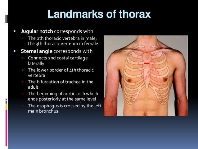
Surface anatomy from image.slidesharecdn.com The 2nd rib articulates on the either side together with the sternum at this level. The rib cage surrounds the lungs and the heart, serving as an important means of bony protection for these vital organs. The junction between the body of the sternum and the xiphoid process is on the level of the tenth thoracic vertebra. The lung is located deep to the area going from axilla to the level of the 7th or 8th rib. It is located at the level of intervertebral disc between the t4 and t5 vertebrae. The typical rib consists of a head neck and body. The neck contains no bony prominences, but simply connects the head with the body. Lateral view of a pair of ribs articulating with the thoracic vertebrae.
Over this surface, except where covered by the scapula, the individual ribs can be distinguished.
An articular capsule surrounds the head of each rib and is further. There are 12 pairs of ribs which are separated by intercostal spaces. 1) the anterior view of the chest wall 2) their relationships to the ribs 3) with the heart superimposed. Because rib 1 articulates with first thoracic vertebra only, there is a single facet on its head (typical ribs have two facets, as mentioned above). The head is wedge shaped, and has two articular facets separated by a wedge of bone. Size of body l3 c2 apex of co. Retrospective assessment of hrct examinations of 250 patients was performed. The localization of the oblique fissures was marked at three site … Free for commercial use no attribution required high quality images. Count the ribs and intercostal spaces. With the upper ribs, closer to the nodule (and in the case of lower ribs, a little further from the nodule) they are curved and have a rough surface that connects them with muscles, angulus costae. Contributing to their role in protecting the internal thoracic organs. Surface markings of the abdomen bony landmarks.—above, the chief bony markings are the xiphoid process, the lower six costal cartilages, and the anterior ends of the lower six ribs.
Therefore it utilized as surface landmark for counting the ribs. The localization of the oblique fissures was marked at three site … There are twelve pairs of ribs, all of which articulate with the vertebral column. The anatomy of the human ribs is made up of 24 ribs which are parted in 12 pairs (each on the left and right side of the chest wall), with the sternum, metasternum (the xiphoid process), and the costal cartilages all situated at the anterior of the chest wall, followed by the thoracic vertebrae on the posterior of the chest wall. Introduction to the structure of the ribcage and ribs:
موقع الدكتور أحمد كلحى: صور تشريح - Anatomy : Thorax ... from 3.bp.blogspot.com Retrospective assessment of hrct examinations of 250 patients was performed. Count the ribs and intercostal spaces. The apex lies above the first rib. Rib 1 is usually shorter and wider than all other ribs, and its broad, flat surface contains grooves that support the subclavian vessels. The uppermost visible digitation of serratus anterior indicates the sixth rib. 1) the anterior view of the chest wall 2) their relationships to the ribs 3) with the heart superimposed. It is an atypical rib and is an important anatomical landmark. A thorough knowledge of thoracic anatomy is of fundamental importance to the thoracic surgeon.
Retrospective assessment of hrct examinations of 250 patients was performed.
Retrospective assessment of hrct examinations of 250 patients was performed. A rib has a flat body, as you can see from the picture of the anatomy of the human rib cage. It is an atypical rib and is an important anatomical landmark. 1) the anterior view of the chest wall 2) their relationships to the ribs 3) with the heart superimposed. The ribs form the main structure of the thoracic cage that protects the thoracic organs. The typical ribs have a generalised structure while the atypical ribs have variations on this structure. Introduction to the structure of the ribcage and ribs: The lung is located deep to the area going from axilla to the level of the 7th or 8th rib. Therefore it utilized as surface landmark for counting the ribs. The first seven sets of ribs, known as true ribs also known as vertebrosternal ribs, are directly articulate with the vertebral column posteriorly and terminate anteriorly as costal cartilage. Anatomy of the human body. The typical rib consists of a head, neck and body: All ribs are attached posteriorly to the thoracic vertebrae.
A rib has a flat body, as you can see from the picture of the anatomy of the human rib cage anatomy of ribs. Therefore it utilized as surface landmark for counting the ribs.